Although pregnancy rates with In Vitro Fertilization have improved drastically in the past decade, many couples using IVF can still fail to achieve a pregnancy. Even those with an excellent prognosis can find it difficult to conceive. This has prompted reproductive specialists to continue looking into ways to improve the treatment.
Several improvements have already been made. This includes the standardization of lab techniques and equipment, and the use of embryo grading. However, even the embryos which have shown the most promise do not always result in pregnancy. Searching for a more effective solution, researchers have now been looking into “Time Lapse Microscopy” (TLM). Here, we will look at how TLM could be the key to improving IVF pregnancy rates.
What is Time Lapse Microscopy?
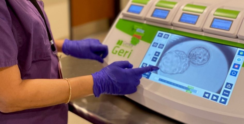
An incubator capable of TLM can capture digital images of embryos during their development in the laboratory. Traditionally, embryologists monitor embryos within the IVF lab at set intervals during the three to five days that the embryos are developing in the lab. They then establish which embryos fail to develop or are slower to develop. This would indicate they would be less suitable for implantation than an embryo that progresses at the expected rate.
TLM is able to monitor the embryos in more regular intervals, without compromising their development. The special incubators contain image-capturing software and a microscope which can capture images every 5-20 minutes.
How can it help with IVF?
Compared to standard incubators, TLM incubators provide a more stable environment. The embryos require a lot less manipulation, which means they are provided with an optimal development environment. Images produced by the special incubators can be assessed by embryologists and physicians both retrospectively and in real-time. This helps them to determine which embryos are better to transfer.
Research is currently being focused on using the data collected from TLM to improve pregnancy rates. The hope is to create an algorithm which would identify the likelihood of the embryo developing through to the blastocyst stage, being chromosomally normal and successful implantating.
In terms of the importance of embryos developing through until the blastocyst stage, research has been somewhat contradictory. Some researchers have discovered that early cell division within the embryo is important; while others have identified later stage division as important. Some studies have also shown that even if an embryo is identified to make it to the blastocyst stage, it did not necessarily improve pregnancy rates.
So, while TLM is an exciting and potentially useful development in IVF, more research is required to determine how it can improve pregnancy rates. The ultimate goal is to create an algorithm which can predict the likelihood of pregnancy based upon the embryos’ development pattern.
Time Lapse Microscopy at GENESIS
The GENESIS IVF laboratory is equipped with time-lapse incubation system. We are able to monitor the development of embryos in order to select the best one for transfer. It is particularly useful for patients who have multiple embryos that are “scored” well based on the standardized grading system. We are able to review the embryo development from day 0 to day 5 and select the one embryo that has clearly demonstrated the most normal development from the beginning.
If you would like to learn more about GENESIS Fertility New York or are ready to schedule an appointment, please speak with one of our representatives at 929-605-5467.